
Why they’re difficult to diagnose and how MRI changes the outcome
Has your horse been “not quite right” for a while? A bit unlevel on a circle? Resistant in collection? Or maybe just not performing the way you know they can? It might be more than a training issue or a bad back. A proximal suspensory injury is one possible cause that can sometimes be challenging to diagnose.
Proximal suspensory injuries are a common cause of lameness and poor performance in horses. Clinical signs can vary depending on whether the forelimbs or hindlimbs are affected. A thorough lameness examination is essential to localise the source of pain. Once the region has been localised, radiography and ultrasonography are typically used to assess the area. Although these techniques can provide important information about both the bone and ligament, each has limitations that can make full assessment of the injury challenging.
What is the proximal suspensory?
The proximal suspensory ligament originates at the top of the cannon bone (Fig 1) and forms the upper portion of the suspensory ligament, a structure that extends down the back of the limb between the splint bones before dividing into branches that attach to the proximal sesamoid bones. The proximal region contains ligamentous tissue interspersed with muscle and fat, and injury may involve the ligament itself, the adjacent bone at its origin, or both.
In some cases, pathology may also affect the branches or their attachment to the proximal sesamoid bones. This is one reason why a thorough assessment of the area is often required to fully understand the nature and extent of an injury.
Injuries affecting the proximal aspect of the suspensory ligament are commonly referred to as proximal suspensory desmitis (PSD) or proximal suspensory disease. They can affect either the forelimbs or hindlimbs and the clinical signs may vary depending on the limb involved and the severity of the injury.

Why is it so hard to spot?
Several things make proximal suspensory injuries difficult to recognise, even for experienced vets.
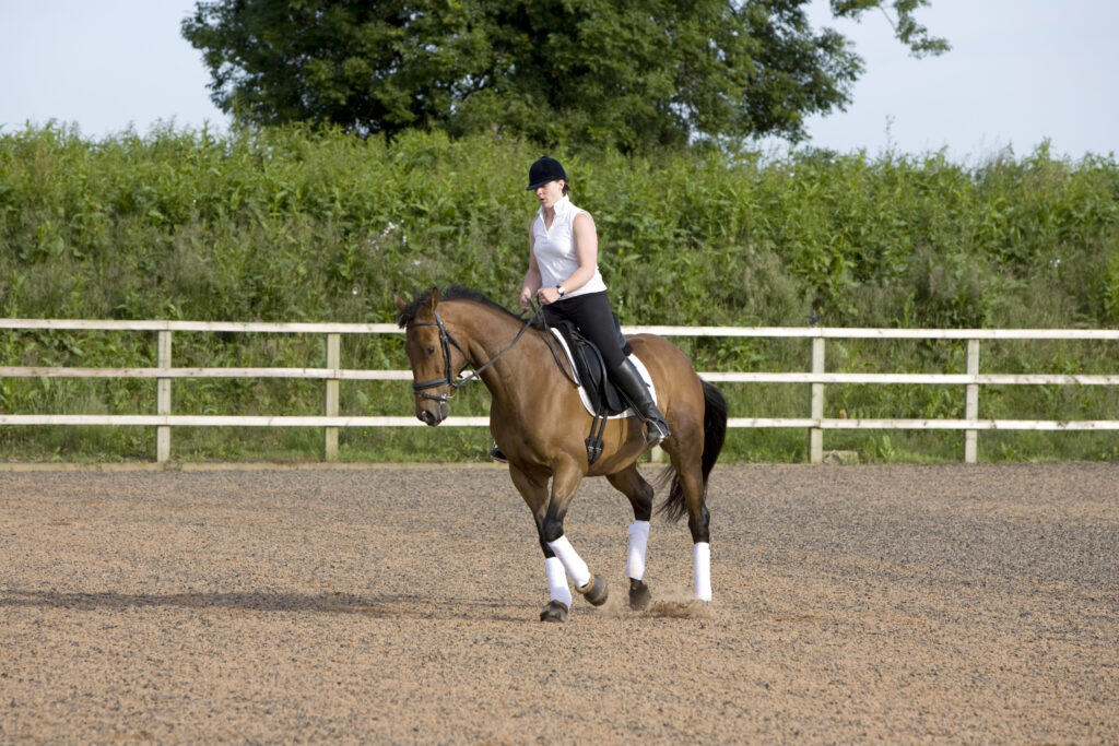
The lameness is often subtle and inconsistent. Your horse might look perfectly fine on a straight line but show discomfort on a circle, particularly on one rein. They might warm up out of it, seeming better after 20 minutes of work rather than worse. This pattern can make owners (and vets) question whether they’re dealing with lameness at all or something more like stiffness or a training problem.
When both hindlimbs are affected, the picture changes again. A horse that is equally uncomfortable on both sides can look remarkably level, because there’s no dramatic contrast between the two limbs. Instead, you might notice a shortened stride, a reluctance to engage behind, a loss of impulsion or power or a general flattening of performance that’s hard to put your finger on.
If you’ve noticed any of these things in your horse, our article Why lameness isn’t always obvious explains why subtle signs matter just as much as dramatic ones.
What does a vet workup involve?
Your vet will watch your horse move – in hand, on a lunge, on different surfaces and in different directions – to build a picture of where and when the discomfort appears. They may use nerve blocks (local anaesthetic injections) to systematically narrow down the source of pain.
This process is explained well in our webinar The value of nerve blocks in the lameness examination.
What’s the challenge with proximal suspensory injuries?
The challenge with proximal suspensory injuries is that confirming the source of pain is not always straightforward. Diagnostic analgesia (nerve blocks) is often used to help localise lameness, but several techniques are available for assessing the proximal suspensory region, each with its own advantages and limitations. In some cases, local anaesthetic may affect adjacent structures, such as the carpal or tarsal joints, which can also be sources of pain, making interpretation of the results more challenging. For this reason, diagnostic analgesia is interpreted alongside the clinical examination and lameness assessment.
Once the proximal suspensory region has been identified as the likely source of pain, radiography and ultrasonography are typically used to investigate the area further. While these modalities provide valuable information about the bone and soft tissue structures, confirming the full extent of the injury can sometimes remain challenging.
Radiography and ultrasonography provide complementary information about the proximal suspensory region:
- Radiographs help evaluate the bone and the ligament’s attachment to it
- Ultrasound assesses the structure and appearance of the ligament itself.
However, some injuries may not be fully characterised using these techniques alone, particularly when determining the extent of bony involvement or whether imaging abnormalities represent active disease. This is where advanced imaging modalities such as MRI can provide additional information.
Why might nothing show up on standard imaging?
This can be one of the most challenging aspects of investigating proximal suspensory injuries. A horse may have clinical signs and diagnostic findings that raise suspicion of proximal suspensory disease, yet radiography and ultrasonography do not always provide a definitive explanation for the lameness or poor performance.
MRI offers a more comprehensive assessment of the region by evaluating both the suspensory ligament and the adjacent bone. It can help identify the location and extent of injury, assess structures that cannot be fully evaluated using other imaging modalities, and provide information that may influence treatment, rehabilitation and prognosis.
“Diagnosing injuries of the proximal suspensory has always proved tricky! But with the right imaging, that no longer has to be the case. Standing MRI delivers a full picture so that a targeted, confident recovery plan for your horse can be put into place.”
Carina Northern, BVSc MRCVS, Vet Surgeon
For a clear explanation of how MRI works and what it can show, take a look at our article: Imaging explained: what is MRI?
What does MRI find that other imaging can’t?
One of the key advantages of MRI is its ability to identify changes within the bone as well as the suspensory ligament. MRI can detect bone oedema, a sign of active bone stress or inflammation that cannot be identified using radiography or ultrasonography. In horses with proximal suspensory disease, pathology may involve the ligament, the adjacent bone, or both. Understanding the extent of bony involvement can be important when developing an appropriate treatment and rehabilitation plan and when discussing prognosis.
MRI can also characterise the ligament injury in detail, such as how much of the ligament is affected, how severe the damage is and whether changes at the attachment point are contributing to the problem. This information shapes the rehabilitation programme: how long, how carefully, and how to monitor progress.
Importantly, standing MRI (where the horse is gently sedated rather than put under general anaesthetic) has become much more effective at imaging the proximal limb. New technology called iNAV improves image quality even when the horse is moving slightly, which was previously a significant challenge for scanning higher up the leg.
To understand what the MRI scan process actually looks like, watch our video The standing MRI process. It takes you through exactly what happens on the day.
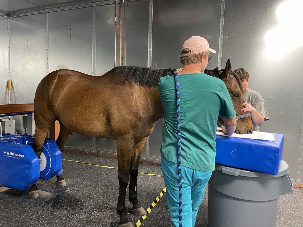
(photo credit: The World Equestrian Centre)
What should you do if you suspect a proximal suspensory injury?
If your horse has been showing subtle, inconsistent lameness, particularly in the hindlimbs, on circles or in collected work, it’s worth asking your vet to discuss whether a full lameness workup is appropriate and whether MRI might be the right next step if standard imaging doesn’t give a clear answer.
You can prepare for that conversation using the Talk Lameness guide: 5 things to discuss with your vet
And you can read more about why getting a diagnosis matters – not just for knowing what’s wrong, but how to fix it – in Lameness diagnosis: why it’s important for horse and owner.
The proximal suspensory is a tricky area. But it’s no longer one that has to stay undiagnosed. With the right imaging, the full picture is now within reach and a targeted, confident recovery plan is the result.