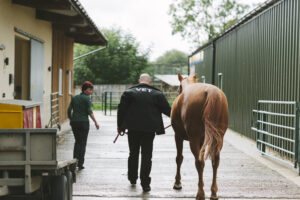
Resources
Check out our resources for practical help and advice on common lameness problems and how best to deal with them.

5 Things to Discuss with Your Vet
In 5 things to discuss with your vet, we help prepare you for a conversation about your horse's lameness. Noting your observations and listing any concerns - in advance of your visit - will be invaluable in helping your vet reach a diagnosis.

Imaging explained: what is MRI?
Magnetic Resonance Imaging (MRI) is an advanced, non-invasive imaging modality. Considered the gold standard for assessing soft tissue injuries, it is also extremely useful for identifying bone pathology, particularly early change which can be missed on X-ray.

First aid for lameness: water & ice
Learning some basic fist aid for lameness will help make a meaningful difference to your horse's recovery. Whether you’re cooling a swollen limb, applying a poultice to a painful foot, or managing a wound with care, these simple first‑aid skills help to ease your horse's discomfort and gather the information your vet needs to make the best decisions.

First aid for lameneness: applying a poultice
Learning some basic fist aid for lameness - like applying a poultice - will help make a meaningful difference to your horse’s recovery. Whether you’re cooling a swollen limb, or managing a wound with care, these simple first‑aid skills help to ease your horse’s discomfort and help you gather the information your vet needs to make the best decisions.

First aid for lameness: wound care
First aid for lameness should include some basic knowledge about wound care. Wounds can be stressful - they look dramatic and horse’s legs are not forgiving places to heal. The good news is that first aid is mostly about three things: safety, cleanliness and knowing if and when you need a vet.

Imaging explained: what is X-ray ?
X-ray, or digital radiography, is one of the primary imaging modalities, mainly used for assessing bone injuries detecting things like fractures, arthritis or bone ‘chips’. X-rays provide limited information on soft tissues such as tendons and ligaments, so vets often pair them with ultrasound or, if needed, advanced imaging. It’s a step by step process!

Imaging explained: what is ultrasound?
Ultrasonography – or ultrasound - is the complementary tool, widely used to assess soft tissue structures such as tendons and ligaments. Like radiography (X-ray), it is portable and generally affordable. However, sound waves cannot easily pass through bone or air, limiting its view to structures adjacent to the bone surface. Ultrasound also allows dynamic assessment of soft tissues when needed.

Imaging explained: what is scintigraphy?
Nuclear Scintigraphy, or a ‘bone scan’, is a technique that assesses physiological processes rather than the anatomy of the organs detecting areas of increased bone remodelling or inflammation ("hot spots"). It is primarily used for investigating musculoskeletal disorders, particularly bone pathology, using an injected radiopharmaceutical which accumulate in areas of bone turnover and increased blood flow (“hot spots”). This modality is sensitive, useful for locating issues like early stress fractures and enthesopathies. However, as it assesses physiological activity rather than anatomical detail, it should not be used in isolation and must be interpreted alongside clinical findings and other imaging techniques. It is also non-portable and requires hospitalisation until radiation levels drop.

Imaging explained: what is CT?
CT (Computed Tomography) is an advanced X-ray–based imaging modality that produces high-resolution, cross-sectional and three-dimensional images. It provides excellent detail of bone and mineralised tissues, particularly in complex anatomical regions such as the head, neck, pelvis and distal limb. With the use of injected contrast, soft tissue structures can also be assessed. CT is fast, typically generating images in seconds to minutes. It is non-portable and - historically - required general anaesthesia, but standing CT systems are now increasingly available.
Imaging explained: what is PET?
PET is the newest advanced functional tool for use in lameness in horses and provides highly sensitive three-dimensional information on tissue metabolic activity. It is particularly useful for detecting areas of increased bone turnover and therefore help distinguish active from inactive lesions. Modern equine PET systems are adapted for standing imaging of the distal limb under light sedation. Increasingly, PET is being used alongside other imaging modalities such as MRI and CT.
