Imaging Explained explores the different modalities available to help with lameness diagnosis, and what you can expect from each. Let’s take a look at scintigraphy or ‘bone scan.’
What is scintigraphy?
Nuclear Scintigraphy, or a ‘bone scan’, is a technique that assesses physiological processes rather than the anatomy of the organs detecting areas of increased bone remodelling or inflammation (“hot spots”). It is primarily used for investigating musculoskeletal disorders, particularly bone pathology, using an injected radiopharmaceutical which accumulate in areas of bone turnover and increased blood flow (“hot spots”). This modality is sensitive, useful for locating issues like early stress fractures and enthesopathies. However, as it assesses physiological activity rather than anatomical detail, it should not be used in isolation and must be interpreted alongside clinical findings and other imaging techniques. It is also non-portable and requires hospitalisation until radiation levels drop.
Your horse will receive a radiopharmaceutical, consisting of a radioactive tracer combined with a pharmaceutical that targets bone, that travels in the bloodstream and accumulates in areas of increased bone turnover or blood flow. The radioactive tracer emits energy that is detected by a gamma camera, highlighting “hot spots” and helping to localise areas of concern, particularly in regions such as the back or pelvis or when multiple limbs are involved. As this technique assesses physiological activity rather than detailed anatomy, findings are interpreted alongside clinical examination and often followed by targeted X-rays, ultrasound or advanced imaging.
When might scintigraphy be used?
- Poor performance or reluctance to work/engage
- Subtle, shifting or multi-limb lameness that’s hard to localise
- Chronic lameness with negative or inconclusive clinical examination, diagnostic analgesia, and radiographic findings
- Cases not amenable to nerve/joint blocks
- Acute severe lameness with no clear findings on clinical examination or initial imaging (radiography/ultrasound)
- Suspected pelvic, spinal, or proximal limb pathology not well assessed with standard imaging
What scintigraphy doesn’t show well
- osteoarthritis with the exception of the hock
- low grade joint disease
- lesions of the coffin and pastern joint
- navicular disease
- OCD lesions, although it can be useful for the stifle and shoulder
- Traumatic fractures if they are scanned shortly after injury
- soft tissue injuries are more difficult to assess
What to expect from scintigraphy
Most horses are scanned standing with mild sedation and the imaging process can take a couple of hours. Because a radioactive tracer is used, short term stable precautions are followed until the tracer decays to safe levels before your horse can return home (usually around 2 days).
Where it fits into the bigger picture
Scintigraphy helps localise areas of concern in complex or poorly localised lameness cases, guiding more targeted imaging such as X-rays, ultrasound, CT or MRI for a definitive diagnosis.
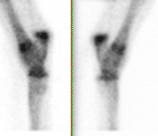
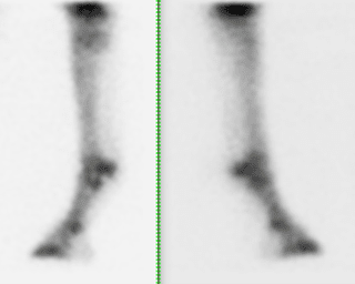
Find out how scintigraphy was used as part of the lameness diagnosis process in a 3-year-old Thoroughbred colt. The patient had pulled up forelimb lame after his last race and subsequently developed a left hindlimb lameness. The cause of the forelimb lameness was identified using diagnostic analgesia and radiography, whilst the hindlimb lameness could not be localised.