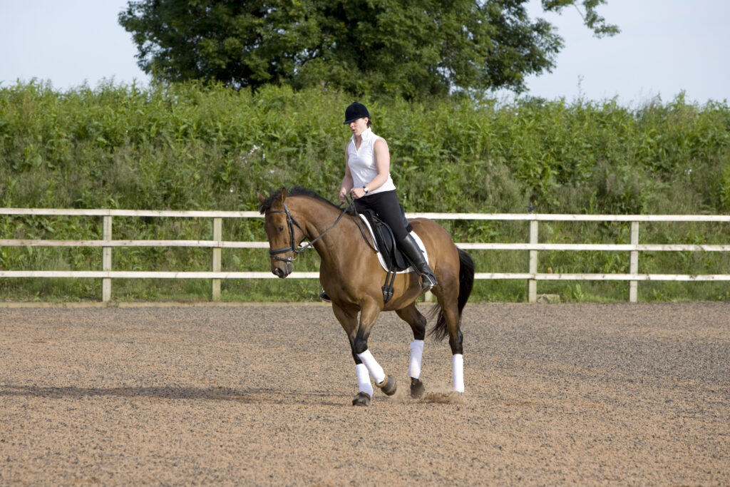
Imaging Explained explores the different modalities available to help with lameness diagnosis and what you can expect from each. We put CT (Computed Tomography) under the spotlight.
What is Computed Tomography (CT)?
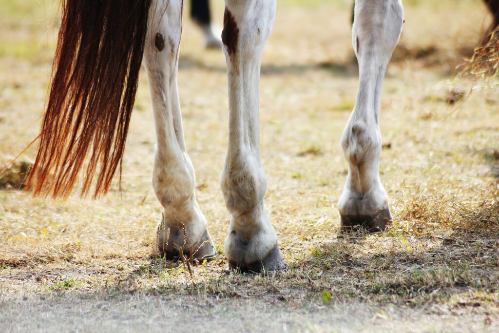
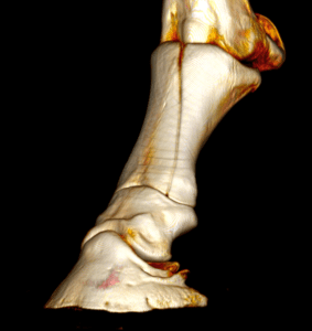
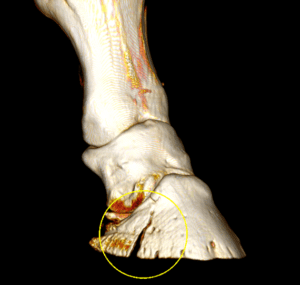
Computed Tomography (CT) is an advanced X-ray–based imaging modality that produces high-resolution, cross-sectional and three-dimensional images. It provides excellent detail of bone and mineralised tissues, particularly in complex anatomical regions such as the head, neck, pelvis and distal limb. With the use of injected contrast, soft tissue structures can also be assessed. CT is fast, typically generating images in seconds to minutes. It is non-portable and – historically – required general anaesthesia, but standing CT systems are now increasingly available.
CT is a 3D imaging technique which uses X-rays. The scanner takes multiple images around the region to be examined, and a computer reconstructs them into high resolution 3D images images. CT is particularly sensitive for detecting structural bone changes, including micro-fractures and variations in bone mineral density, which may be missed on standard radiographs. However, CT does not assess bone oedema which can be an early stage physiological change and is less sensitive than MRI for soft tissue injuries.
When might CT be used?
- When detailed assessment of bone is required, particularly if X-rays are inconclusive
- Investigation of complex bony regions (e.g. foot, fetlock, head/neck, sinuses, teeth, pelvis)
- Detection of subtle or complex fractures, bone lesions, or areas of bone remodelling
- Surgical planning where precise anatomical detail is needed
What it shows well
- Fine bone architecture, including cortical and trabecular bone, joint surfaces, and small fragments (e.g. chips or micro-fractures)
- Structural changes such as sclerosis, bone resorption, and complex fractures
- Three-dimensional relationships of structures without superimposition or ‘overlap’ seen on radiographs
What it doesn’t show well
- Soft tissues can be assessed with the administration of contrast, but not with the detail of MRI
- Some regions still require general anaesthesia depending on system and area scanned; many distal limb studies are possible standing (clinic‑dependent)
What to expect from CT
Your vet will advise whether your horse can be scanned standing (mild sedation) or under general anaesthesia. This decision will be based on the area to be scanned and resources at the facility. The scan itself is quick; preparation and positioning take longer.
Where it fits into the bigger picture
CT is typically chosen when detailed assessment of bone is required and is likely to influence diagnosis or treatment decisions. If soft tissue evaluation is the priority – particularly within the hoof capsule – MRI may be recommended instead.
Interested in CT? Then take a look at The Complete Guide which includes how to prepare your horse for a standing CT scan and walks you through the procedure. It’s available on the link below.