Why they’re difficult to diagnose and how MRI changes the outcome
Has your horse been “not quite right” for a while? A bit unlevel on a circle? Resistant in collection? Or maybe just not performing the way you know they can? It might be more than a training issue or a bad back. A proximal suspensory injury is one possible cause that can sometimes be challenging to diagnose.
Proximal suspensory injuries are a common cause of lameness and poor performance in horses. Clinical signs can vary depending on whether the forelimbs or hindlimbs are affected. A thorough lameness examination is essential to localise the source of pain. Once the region has been localised, radiography and ultrasonography are typically used to assess the area. Although these techniques can provide important information about both the bone and ligament, each has limitations that can make full assessment of the injury challenging.
What is the proximal suspensory?
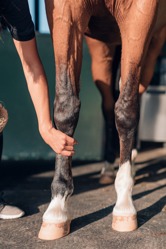
The proximal suspensory ligament originates at the top of the cannon bone (Fig 1) and forms the upper portion of the suspensory ligament, a structure that extends down the back of the limb between the splint bones before dividing into branches that attach to the proximal sesamoid bones. The proximal region contains ligamentous tissue interspersed with muscle and fat, and injury may involve the ligament itself, the adjacent bone at its origin, or both.
In some cases, pathology may also affect the branches or their attachment to the proximal sesamoid bones. This is one reason why a thorough assessment of the area is often required to fully understand the nature and extent of an injury.
Injuries affecting the proximal aspect of the suspensory ligament are commonly referred to as proximal suspensory desmitis (PSD) or proximal suspensory disease. They can affect either the forelimbs or hindlimbs and the clinical signs may vary depending on the limb involved and the severity of the injury.
Fig. 1: The cannon bone is the main load‑bearing bone in the horse’s lower limb, running between the knee and the fetlock
Why is it so hard to spot?
Several things make proximal suspensory injuries difficult to recognise, even for experienced vets.
The lameness is often subtle and inconsistent. Your horse might look perfectly fine on a straight line but show discomfort on a circle, particularly on one rein. They might warm up out of it, seeming better after 20 minutes of work rather than worse. This pattern can make owners (and vets) question whether they’re dealing with lameness at all or something more like stiffness or a training problem.
When both hindlimbs are affected, the picture changes again. A horse that is equally uncomfortable on both sides can look remarkably level, because there’s no dramatic contrast between the two limbs. Instead, you might notice a shortened stride, a reluctance to engage behind, a loss of impulsion or power or a general flattening of performance that’s hard to put your finger on.
If you’ve noticed any of these things in your horse, our article Why lameness isn’t always obvious explains why subtle signs matter just as much as dramatic ones.
What does a vet workup involve?
Your vet will watch your horse move – in hand, on a lunge, on different surfaces and in different directions – to build a picture of where and when the discomfort appears. They may use nerve blocks (local anaesthetic injections) to systematically narrow down the source of pain.
What’s the challenge with proximal suspensory injuries?
The challenge with proximal suspensory injuries is that confirming the source of pain is not always straightforward. Diagnostic analgesia (nerve blocks) is often used to help localise lameness, but several techniques are available for assessing the proximal suspensory region, each with its own advantages and limitations. In some cases, local anaesthetic may affect adjacent structures, such as the carpal or tarsal joints, which can also be sources of pain, making interpretation of the results more challenging. For this reason, diagnostic analgesia is interpreted alongside the clinical examination and lameness assessment.
Once the proximal suspensory region has been identified as the likely source of pain, radiography and ultrasonography are typically used to investigate the area further. While these modalities provide valuable information about the bone and soft tissue structures, confirming the full extent of the injury can sometimes remain challenging.
Radiography and ultrasonography provide complementary information about the proximal suspensory region:
Radiographs help evaluate the bone and the ligament’s attachment to it
Ultrasound assesses the structure and appearance of the ligament itself.
However, some injuries may not be fully characterised using these techniques alone, particularly when determining the extent of bony involvement or whether imaging abnormalities represent active disease. This is where advanced imaging modalities such as MRI can provide additional information.
Why might nothing show up on standard imaging?
This can be one of the most challenging aspects of investigating proximal suspensory injuries. A horse may have clinical signs and diagnostic findings that raise suspicion of proximal suspensory disease, yet radiography and ultrasonography do not always provide a definitive explanation for the lameness or poor performance.
MRI offers a more comprehensive assessment of the region by evaluating both the suspensory ligament and the adjacent bone. It can help identify the location and extent of injury, assess structures that cannot be fully evaluated using other imaging modalities, and provide information that may influence treatment, rehabilitation and prognosis.
“Diagnosing injuries of the proximal suspensory has always proved tricky! But with the right imaging, that no longer has to be the case. Standing MRI delivers a full picture so that a targeted, confident recovery plan for your horse can be put into place.”
One of the key advantages of MRI is its ability to identify changes within the bone as well as the suspensory ligament. MRI can detect bone oedema, a sign of active bone stress or inflammation that cannot be identified using radiography or ultrasonography. In horses with proximal suspensory disease, pathology may involve the ligament, the adjacent bone, or both. Understanding the extent of bony involvement can be important when developing an appropriate treatment and rehabilitation plan and when discussing prognosis.
MRI can also characterise the ligament injury in detail, such as how much of the ligament is affected, how severe the damage is and whether changes at the attachment point are contributing to the problem. This information shapes the rehabilitation programme: how long, how carefully, and how to monitor progress.
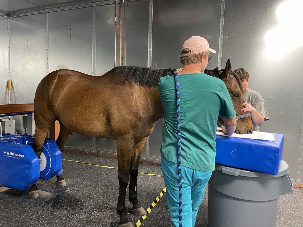
Importantly, standing MRI (where the horse is gently sedated rather than put under general anaesthetic) has become much more effective at imaging the proximal limb. New technology called iNAV improves image quality even when the horse is moving slightly, which was previously a significant challenge for scanning higher up the leg.
To understand what the MRI scan process actually looks like, watch our video The standing MRI process. It takes you through exactly what happens on the day.
Standing MRI has become much more effective at scanning the proximal limb (photo credit: The World Equestrian Centre)
What should you do if you suspect a proximal suspensory injury?
If your horse has been showing subtle, inconsistent lameness, particularly in the hindlimbs, on circles or in collected work, it’s worth asking your vet to discuss whether a full lameness workup is appropriate and whether MRI might be the right next step if standard imaging doesn’t give a clear answer.
The proximal suspensory is a tricky area. But it’s no longer one that has to stay undiagnosed. With the right imaging, the full picture is now within reach and a targeted, confident recovery plan is the result.
There’s a particular feeling that arrives in the spring: the mornings get lighter, the yard smells of wet earth and new grass and suddenly you want to do more with your horse. After months of short days, heavy going and limited riding, spring feels like permission to start again.
But horses don’t reset overnight. Their bodies have been managing winter – lower workloads, different feed, heavier rugs, harder or muddier ground, less varied movement. As the season shifts and your ambitions grow, it’s worth pausing before you push forward. A little attention now across a handful of areas can make the difference between a horse that builds through spring and one that struggles into summer.
This isn’t a list of problems to worry about. It’s a prompt to look carefully at your horse before the season gets away from you.
The ground is changing – and so is the risk
One of the most significant shifts in spring is one you might not immediately connect to your horse’s wellbeing: the grass.
After a winter of slow growth, spring grass comes in fast and rich, packed with non-structural carbohydrates – the sugars and starches that accumulate as plants photosynthesise in longer daylight hours. For most horses this isn’t a crisis. But for those prone to laminitis, insulin resistance or carrying extra condition through winter, spring grass is the biggest risk they’ll face all year.
The signs of early laminitis can be easy to miss. A horse that seems a little sore, reluctant to step forward or shifting weight more than usual may be telling you something important. Feet that feel warmer than normal, a stronger digital pulse or reluctance to trot on hard ground are all worth noting.
If your horse is in this category, gradual introduction to pasture makes a meaningful difference. Short initial turnout (even just 15 to 20 minutes) with gradual increases over several weeks, is far better than suddenly switching from a hay-based winter diet to unlimited spring grazing. Giving hay before turnout helps reduce the urge to gorge. And if you’re turning out on lush grass, earlier morning or overnight grazing tends to mean lower sugar levels than afternoon, though this isn’t foolproof in spring conditions.
If something feels off, trust that instinct. The Talk Lamness Quiz is a good starting point for thinking through what you’re seeing and how to describe it to your vet.
Feed: the transition matters as much as the destination
A horse’s digestive system adapts to its diet over time and the microbiome in the hindgut, which does so much of the work of processing forage, doesn’t appreciate sudden change. If you’re moving from a winter feed regime to something richer, or increasing calories to build condition ahead of a competition season, do it slowly.
As a rule of thumb, make any significant feed change over at least two weeks, ideally longer. Monitor your horse’s weight and condition as you go. What looks like “catching up” after winter can tip into overweight quite quickly in spring, particularly in good doers and native breeds.
If your horse has been stabled for much of the winter, digestive changes – including an increased risk of colic – can be associated with the transition back to more grass. Keep an eye on gut sounds, droppings and general demeanour as turnout increases.
Fitness: patience pays
After a winter of reduced or light work, a horse’s cardiovascular fitness, muscle strength and the integrity of tendons and ligaments will all have dropped. Connective tissue in particular responds slowly to training load – it strengthens, but over months rather than weeks.
If you have events or competitions in mind for the season ahead, map out a realistic timeline. Six to eight weeks of gradually increasing work before you ask for anything demanding is a minimum and that’s for a horse that’s been in light work through winter. A horse that’s had complete rest needs longer.
The classic mistake in spring is doing too much too soon in the enthusiasm of better weather and firmer going. Soft tissue injuries, like suspensory strains or tendon issues, are disproportionately common at this time of year and they’re the kind of setbacks that cost months, not weeks.
Build base fitness before you add speed, intensity or technical demands. Hacking on varied terrain, long and low schooling and steady hill work are all excellent early season tools. Save the harder stuff until your horse is clearly moving freely and willingly.
Watch how your horse moves. Are they tracking up evenly? Is there any shortening of stride, reluctance on one rein or resistance in transitions that wasn’t there before? These can be early signs of discomfort that are much easier to address if caught early.
A horse that isn’t quite right in spring , even subtly, is telling you something before it becomes a problem you can’t ignore.
Many horses spend winter in rugs that are heavier than they strictly need and many come off rugs in spring either too abruptly or later than is comfortable. Horses regulate their temperature remarkably well and a horse in light work – that’s been trace or full clipped through winter – may be genuinely ready to come out of a heavyweight rug sooner than you think once temperatures stabilise.
Check the fit of rugs that have been on all winter. Horses lose and gain condition through winter and a rug that fitted in November may be sitting differently by May. Look for signs of rubbing at the shoulders, withers and chest, which can indicate poor fit and cause low-grade discomfort that’s easy to overlook.
A horse that’s been rugged heavily through a mild winter may also be carrying more condition than you realise. The rug can mask a rounder topline and ribs that are harder to feel than they should be. Spring is a good moment to take the rug off, step back and actually look at your horse’s body condition honestly.
Tack fit: a winter body is not a spring body!
This is one of the most commonly overlooked areas of spring management. Horses change shape through winter (you may well know that feeling yourself as you start to shed those winter jumpers!). Some lose muscle and topline, others gain weight – and a saddle might not sit the same by spring.
A saddle that was well balanced in autumn can appear to tip forward or rock on a horse that’s lost condition through its back, creating pressure points that weren’t there before. Conversely, a horse that’s gained weight across the back and shoulders may suddenly find its saddle feels tight in ways it didn’t.
If you’re planning to increase work significantly in spring, a saddle fit check before the season starts, not after a problem appears, is money well spent. The same applies to bridle fit, particularly if you use anatomical or shaped bridles where the padding placement matters.
Feet: hoof growth picks up in spring
Hooves grow faster in spring as the ground softens and nutrition improves. For horses in work, this means your farrier schedule may need revisiting. A horse that was comfortable on a 10-to-12-week interval through winter may start to go long in the toe or lose balance in the hoof capsule more quickly once spring kicks in.
Subtle foot pain is one of the most common forms of lameness in horses so early investigation pays dividends. A horse that’s mildly foot sore in spring can deteriorate quickly once the workload increases.
If your horse is barefoot, keep a close eye on wear versus growth. Spring going can be unpredictably variable, and a horse that was managing fine on hard winter ground may need more frequent attention as the going changes.
For all horses, spring is a natural time to discuss with your farrier whether the previous season’s approach is still right. Changes in workload, surfaces or the horse’s way of going can all influence the best approach to shoeing or trimming. If your horse is showing any foot soreness, even mild or intermittent, don’t assume it’s just the changing ground. Subtle foot pain is one of the most common forms of lameness in horses and it’s one of the areas where early investigation pays dividends. A horse that’s mildly foot sore in spring can deteriorate quickly once the workload increases.
Final thoughts: use spring as an opportunity to observe
The seasonal change is one of the best prompts to really look at your horse: not with a list of concerns in mind, but just with fresh eyes. Watch them move in the field. Feel along their back and legs. Notice if their attitude to work is different. Has anything changed since autumn?
Horses are very good at compensating for low-grade discomfort, which is why problems can become established before they become obvious. The patterns you notice over a few days – a slight reluctance on the right rein, a change in behaviour when you groom over the back, a foot that’s warmer than the others – are often more useful information than a single observation.
Spring is also an excellent time to raise anything you’ve noticed with your vet, before the season gets into full swing and your diary fills up. If you’re not sure how to describe what you’re seeing, or whether it warrants a call, 5 things to discuss with your vet is a practical guide to preparing for exactly that conversation.
If your horse is showing signs of lameness — or something that’s just not quite right — Talk Lameness is a free educational resource to help you understand what you’re seeing and talk confidently with your vet. Start the Quiz.
For equine vets and practices interested in standing MRI for lameness diagnosis, find out more about Hallmarq’s equine imaging. With the tools available to scan the foot, carpus and tarsus, the gold standard in lameness diagnosis is now even better!
If you’ve ever thought “something’s off with my horse, but I can’t prove it”, you’re not imagining things. Lameness isn’t always a dramatic limp. More often, it’s a collection of small changes that come and go, shift with the surface or only show up when you’re riding.
When vets measure lameness, they’re doing one main thing: turning a gut feeling into a clear picture they can track, compare and use to decide what to do next. It’s not usually one magic number. It’s a story built from a few reliable clues.
1. First, we agree what we’re looking at
In plain terms, lameness is a change in the way a horse moves that’s linked to discomfort or reduced function. The tricky bit is that horses are brilliant at coping. They can shift weight, shorten a stride, change rhythm or alter balance so the problem hides in plain sight.
That’s why measuring lameness is less about catching one bad step and more about noticing patterns and unevenness. It’s asking: is something consistently different on one side, under certain conditions or over time?
2. The trained eye: what your vet is watching for
A lameness assessment usually starts with careful observation at walk and trot, often on a firm, level surface. Your vet is looking at stride length, rhythm, straightness and how evenly your horse loads and pushes off each limb. They’ll often watch from the side and from behind, because different angles show different clues.
Then they’ll add context. That might mean circles, different surfaces or seeing the horse under saddle if that’s where the issue shows up. The point isn’t to make the horse look worse. It’s to create the conditions where subtle issues are easier to spot, so your vet can start narrowing things down.
“Measuring lameness isn’t about proving anything. It’s about building a clearer picture so you and your vet can make better decisions. The better the picture, the calmer the plan tends to be.”
Chrysanthi Pitaouli, DVM, CertAVP
3. Grading scales: a shared language, not a label
To make lameness easier to describe, vets often use a grading scale. You might hear the AAEP 0–5 scale mentioned, where 0 is not perceptible and 5 is non weight bearing. The middle grades are where many “not quite right” horses sit. They might look fine some of the time, then show it on a circle, on a hard surface, when they’re tired or when they’re ridden.
A grade isn’t a judgement on you or your horse. It’s simply a way of saying “today, in these conditions, this is how clear it is”, so it can be compared later.
4. Making it repeatable: conditions matter
One reason lameness feels so confusing is that it can change depending on:
the surface (hard vs soft)
straight lines vs circles
warming up vs fatigue
ridden vs in-hand
So part of measuring lameness is keeping things consistent. Vets do this by repeating the same checks and comparing what they see in different situations.
As an owner, you don’t need to recreate a full work-up at home. But you can help by keeping your horse’s story tidy. Two short videos filmed in a similar way a few days apart, plus a note of when it looks better or worse, can be genuinely useful.
5. Objective gait analysis: when sensors help
Alongside the vet’s eye, there are tools designed to measure movement more objectively. Some practices may use systems that allow for small sensors to be placed on areas like the poll and pelvis to measure asymmetry and track change over time.
This can be helpful when:
signs are subtle and you want to be sure a change is real
the picture is complicated (for example, multiple issues or compensation)
your vet wants a consistent way to monitor response to treatment or rehab
It’s worth saying: sensors don’t diagnose the cause on their own. They’re another way of measuring what’s happening, so decisions aren’t based on guesswork.
6. Measuring response: what changes after a test?
A big part of lameness measurement is comparison. Your vet will look at your horse’s movement at baseline, then see what changes after specific steps in the exam.
That might include flexion tests, hoof testing or local anaesthetic blocks used to narrow down where discomfort is coming from. The principle is the same each time: they’re not just looking at what you see, they’re looking at what changes when something is tested.
This step-by-step approach is why lameness work-ups can feel slow. They’re designed to build accuracy, not rush to a conclusion.
7. Imaging: getting a clearer picture inside
Sometimes the lameness is measurable, but the why still isn’t clear. That’s where imaging comes in.
X-rays and ultrasound can be really useful depending on what your vet suspects. Ultrasound is often used to look at soft tissues that are accessible from the outside. X-rays help with bony changes and joint spaces.
MRI is another option, particularly in complex areas like the foot and lower limb, where structures are crowded and harder to fully assess with other imaging. Standing equine MRI can often be done with the horse standing under mild sedation. It’s one tool among several, used when your vet needs more detail to guide treatment, rehab or next steps.
The takeaway
Measuring lameness isn’t about proving anything. It’s about building a clearer picture so you and your vet can make better decisions. The better the picture, the calmer the plan tends to be.
If you want a practical next step, keep it simple:
film a short straight line trot clip if it’s safe
jot down when it looks worse or better (surface, rein, warm-up, ridden vs in-hand)
share that with your vet
You’re not expected to have the answers. You’re just helping tell your horse’s story clearly and with confidence!
It’s not just humans that feel more aches and pains as they get older! Your horse may well be showing signs of aging in small and often subtle ways that are all too easy to overlook. When it comes to lameness diagnosis, knowing your horse’s age can help your vet give the right treatment.
Age can help shape the probable causes of lameness and guide the next steps in diagnosis and care. Recognising symptoms and patterns that are normal for your horse and those that signify something more serious or that needs attention, will help you and your vet in getting the right answers, without guesswork.
Why age is such a powerful clue
Lameness isn’t a disease; it’s a sign that something hurts or isn’t working as it should within the musculoskeletal system: hooves, tendons, ligaments, joints, muscles, even the back or neck. The likely culprit shifts with age, and vets use that context to narrow possibilities quickly and choose the most useful and informative tests first.
Older horses: more often show chronic or degenerative issues. For example, arthritis (degenerative joint disease), age‑related tendon or ligament weakening, or PPID (Cushing’s) associated laminitis risk. These problems tend to build gradually, so signs can be subtle: shorter steps, stiffness after rest, reluctance to bend, or a quiet change in demeanour.
Younger horses: are more likely to face acute injuries from training, turnout mishaps or workload changes. Think tendon or ligament strains, bruised soles, or developmental issues. Their onset is often faster, more obvious, and can be managed in the shorter term for an effective outcome.
A better understanding of age related patterns of movement and pain-related behaviours will help both you and your vet to move with purpose rather than uncertainty.
Lameness isn’t a disease; it’s a sign that something hurts or isn’t working as it should.
Older vs younger: what vets expect (and why it matters)
In older horses
Lameness is often chronic or stress‑related with wear‑and‑tear that has accumulated over the years. Vets commonly look for:
Arthritis and joint effusion (extra joint fluid), sometimes presenting first as mild stiffness or a shorter stride.
Age‑related tendon/ligament degeneration that can flare with apparently minor effort or uneven footing.
Endocrine links, especially PPID, which heightens the risk of laminitis and the “short, tentative” steps owners often describe.
Because changes are gradual, daily observation – how your horse stands, turns, and moves – becomes your strongest asset. Simple limb palpation and knowing your horse’s “normal” movement will help you any spot new swelling or heat early.
Ipsum sit mattis nulla quam nulla. Gravida id gravida ac enim mauris id. Non pellentesque congue eget consectetur turpis. Sapien, dictum molestie sem tempor. Diam elit, orci, tincidunt aenean tempus. Quis velit eget ut tortor tellus. Sed vel, congue felis elit erat nam nibh orci.
“Your horse’s age is not just a number: it’s a diagnostic advantage.
”
Younger horses typically need a repair‑and‑rehabilitaiton plan designed for full recovery:
Rest and progressive loading for soft‑tissue injuries, based on imaging findings.
Targeted physiotherapy/exercise prescription and changes to work or surfaces to prevent recurrence.
In both older and younger horses, a clear diagnosis prevents you from trying lots of small changes without knowing what helps. The plan can be targeted for effective results and the quickest possible best outcome for your horse.
What you can do right now (age-aware tips)
Watch and observe your horse’s daily patterns and movements. Make notes so that you can tell your horse’s lameness story:
Notice the small things: daily checks of posture, weight‑bearing, and mood often flag change earlier than gait alone, especially in more senior horses.
Watch in motion: a brief trot‑up on a straight line and on a circle (both reins) can reveal head‑bob (front legs) or hip asymmetry (hind legs). Any video footage of this trot-up will help you and your vet review subtle patterns.
Use your hands: run your hands over your horse’s legs and compare limbs for heat, swelling or sensitivity. Knowing your horse’s “normal” makes any new changes more obvious.
Share specifics with your vet: age, onset (sudden vs gradual), any training/surface/shoeing changes, and what you’ve already tried all speed up the route to the right treatment.
The takeaway
Your horse’s age is not just a number: it’s a diagnostic advantage.
For older horses, think chronic or degenerative patterns that benefit from thoughtful, long‑term management. For younger horses, think acute injury with focused imaging and rehab. Either way, your careful observations plus your vet’s age‑informed approach will lead to a clearer diagnosis and the right treatment, faster.
Sources & further reading
Owner‑friendly overview of lameness, causes and the value of a structured exam (MSD/Merck Veterinary Manual). [msdvetmanual.com]
Behavioural indicators and recognition of subtle lameness signs (British Horse Society; Horses Inside Out). [bhs.org.uk], [horsesinsideout.com]
Professional outline of the lameness examination, from history to imaging. [msdvetmanual.com]
Footnote
Lameness diagnosis is important for both you and your horse. As an owner, recognising and diagnosing lameness is crucial to ensuring your horse’s long-term soundness. Early diagnosis provides clarity, reduces stress, and helps you and your vet to make informed decisions to support your horse’s health.
Myths about horse lameness abound, especially when people are not armed with the facts. If lameness were always obvious, none of us would spend weeks second guessing what we’re feeling under saddle. Lameness can be subtle, inconsistent and frustratingly hard to describe. That is why myths take hold! They give simple answers to something that is rarely simple.
This article is here to gently clear the air. No judgement, no diagnosis, just a clearer way to think about what you are seeing in your horse and what you can do next.
“If lameness were always obvious, none of us would spend weeks second guessing what we’re feeling under saddle. ”
Why people say it: We picture lameness as an obvious head bob or a clear short step.
What is usually truer: Not all lameness looks like a classic limp. Some horses stay straight but feel uneven on one rein, struggle in transitions, lose impulsion or change their way of going. Subtle lameness is often more about patterns than one big moment.
What to do instead: Pick one or two consistent checks you can repeat, like straight lines in trot on a firm, level surface, then a circle on each rein. Keep it calm and brief. If you can, take short videos from the side and behind to share with your vet.
Myth 2 – it’s just behavioural
Why people say it: Behaviour is visible. Pain is not. It is easy to label reluctance as naughtiness, stubbornness or a training issue.
What is usually truer: Discomfort can sometimes look like behaviour. Resistance, crookedness, refusing one lead, sudden loss of confidence or a horse that feels “cold backed” can all be ways of saying something does not feel right. That does not mean every training problem is lameness, but it does mean it is worth staying curious.
What to do instead: Swap blame for observation. What exactly changed? When do you notice it most? Is it worse on a circle, on one rein, on hard ground, or when they are tired? Those details help your vet far more than a label.
Myth 3 – if it comes and goes, it can’t be serious
Why people say it: We expect pain to be constant.
What is usually truer: Lameness can be intermittent. Some horses look better after warming up, then worsen with fatigue. Others are fine on grass but struggle on a firmer surface. Some only show it on the lunge, under saddle or when asked for a certain movement. Intermittent does not mean imaginary. It often means the conditions matter.
What to do instead: Instead of trying to catch it in the act, track the pattern. A simple note on your phone is enough: date, work done, surface, rein, gait and what you noticed.
Myth 4 – a bit of a rest will sort it out
Why people say it: Rest feels kind and sometimes it does help.
What is usually truer: Rest can reduce visible signs, but it does not always answer the why. Some problems improve with time and correct rehab. Others return as soon as work resumes because the underlying issue has not been identified. That is why vets use a step-by-step work-up to narrow down where pain is coming from, then choose treatment and rehab based on evidence.
What to do instead: If you are considering rest, do it with a plan and your vet involved. Ask what they want you to monitor, when to recheck and what the next step would be if it returns. In the first instance, they may suggest X-ray or ultrasound. If these first line imaging options don’t deliver any answers, then advanced imaging such as MRI may well be suggested.
Myth 5 – advanced imaging is expensive and only for the worst cases
Why people say it: Advanced imaging can feel extreme and often requires travel to a vet practice.
What is usually truer: ‘Advanced’ simply means a more complicated piece of equipment is used, which is usually not portable, and therefore travelling to a clinic for a day procedure is common. Advanced imaging can include: Scintigraphy (‘bone scan’), Computed Tomography (CT), or Magnetic Resonance Imaging (MRI), and nearly all can be performed while the horse is awake and under mild sedation.
Advanced imaging techniques are recommended because they enable further information to be obtained about soft tissues, bone changes, or both, when a diagnosis hasn’t been reached yet. Advanced imaging shouldn’t be reserved for the worst cases only, however. Depending on chronicity, it’s only used when a lameness is deemed to be at its worst, very severe, or has been going on for a long time; it may be more difficult to identify the primary cause from issues that have developed as a result of the length of time the lameness has been present. Or even when a diagnosis is made, it may be accompanied by a poor prognosis for recovery because of the delay in starting appropriate treatment.
None of this means advanced imaging is the right step for every horse. It simply means it can be a sensible part of the decision-making journey when you vet needs more detail to guide treatment, rehab, or next steps.
What to do instead: If advanced imaging has been mentioned, ask your vet:
What questions are we trying to answer now?
What have we already ruled out?
Which modality would they suggest and why?
A quick reminder
Talk Lameness is for education, not diagnosis. If you’re worried about your horse, it’s always a good idea to speak to your vet.
Footnote
Lameness diagnosis is important for both you and your horse. As an owner, recognising and diagnosing lameness is crucial to ensuring your horse’s long-term soundness. Early diagnosis provides clarity, reduces stress, and helps you and your vet to make informed decisions to support your horse’s health.
If lameness always looked like a clear limp, most of us would spot it in five minutes, call the vet and move on. In real life, it’s rarely that tidy. More often, you notice a feeling first. Your horse is a touch uneven, a bit reluctant or just not moving like themselves and you start second guessing. Are you imagining it? Is it the surface? Are they being cheeky? Is it your riding?
That uncertainty is exactly why lameness can be so stressful. It’s also why it helps to understand one simple truth: horses don’t all show pain in the same way and movement changes can be subtle long before they’re obvious.
Here’s why.
Horses are built to cope
Horses are prey animals. Their instinct is to keep going, even when something hurts. Many horses will protect a sore area by shifting weight, changing their rhythm or loading a different part of the limb. To you, it can look like a normal day with a few little quirks. To your vet, those quirks can be clues.
This coping is also why you might feel something is off under saddle, even when your horse looks fine to someone watching from the gate.
Subtle lameness doesn’t always look like a limp
A limp is one way lameness can show up, but it’s not the only one. Some horses shorten a stride rather than clearly nod. Some move evenly in a straight line but look different on a circle. Some lose power behind, feel less willing or struggle with a lead that was always easy.
You might notice:
a change in rhythm or regularity at trot
unevenness that comes and goes
reluctance to bend one way
stumbling or toe dragging
difficulty with transitions
a drop in performance, forwardness, or confidence
None of these proves lameness on their own. But they can be the start of a story worth paying attention to.
Horses don’t all show pain in the same way and movement changes can be subtle long before they’re obvious.
“Lameness often shows up under certain conditions, not all the time. That’s why two people can look at the same horse and see different things.”
Humphrey Grimmett BVetMed BSc MRCVS, Vet Surgeon
Lameness can depend on the situation
Lameness often shows up under certain conditions, not all the time. That’s why two people can look at the same horse and see different things.
Common “it only happens when…” moments include:
on a circle, especially on one rein
on a firm surface, or sometimes only on softer ground
when the horse is tired
after warming up, or sometimes only before they’ve loosened up
under saddle, but not obviously in-hand
This isn’t your horse being dramatic. It’s just biomechanics. Different surfaces and shapes change how each limb is loaded and that can make a mild issue easier or harder to see.
Compensation muddies the picture
One sore area can cause knock-on changes elsewhere. A horse protecting a front foot might alter shoulder movement, change how they use their neck or shift weight back. A hindlimb issue can change how the horse carries their pelvis, how they sit in canter or how forward going they are.
This is why lameness isn’t always as simple as “left fore”. Sometimes the visible change is a compensation, not the original source. It’s also why vets work step by step, because they’re trying to find what’s driving the pattern, not just what looks odd.
Behaviour can be part of the lameness story
This is a big one and it’s where owners often feel judged.
Sometimes discomfort shows up as resistance, tension, napping, bucking, refusing or just feeling flat. That doesn’t mean every behaviour issue is pain. But it does mean behaviour doesn’t automatically rule pain out either.
A useful way to think about it is this: behaviour is information. It’s one piece of the picture, alongside movement, context and patterns over time.
Your eye adapts without you realising
If you see your horse every day, tiny changes can creep in gradually. You adjust your expectations without meaning to. It’s why someone who hasn’t seen them in a month might say, “they look different”, and you’re left thinking, do they?
This is also why short videos can be so helpful. They freeze a moment in time. You can compare week to week or show your vet exactly what you’re trying to describe.
What to do if you’re not sure
You don’t need to prove anything. You just need to get curious and collect a few useful details. A simple plan:
Notice the pattern: when is it worse, when is it better, what changed recently
Film a short clip if it’s safe: straight line trot and a circle each rein if you can
Write a few notes: surface, rein, warm-up, ridden vs in-hand
Talk to your vet if you’re worried or if it’s persistent or getting worse
You’re not overreacting by asking questions. You’re doing what good horse people do. You’re paying attention and you’re learning to talk lameness so your horse gets the right help sooner.
Footnote
Lameness diagnosis is important for both you and your horse. As an owner, recognising and diagnosing lameness is crucial to ensuring your horse’s long-term soundness. Early diagnosis provides clarity, reduces stress, and helps you and your vet to make informed decisions to support your horse’s health.
Ever get the feeling that your horse is not “quite right?” Well, the feeling is real, even when you can’t point to one clear moment where they look lame. The goal is not to diagnose at home. It’s to turn a vague worry into a clear, calm set of observations your vet can use.
1. Press pause: take the emotion out of the first check
Before you change everything at once, take a breath and do a quick reset. Ask yourself:
What’s changed in the last 2-3 weeks if anything? Workload, surface, shoeing, turnout, feel, saddle, rider
Is it worse on one rein, on a circle, or a firm surface, or when tired?
Is it new, getting worse, or staying the same?
If your gut says your horse is in pain, or anything looks sudden or severe, call your vet sooner rather than later.
2. Do a simple, repeatable “baseline” check
You’re looking for patterns, not proof. In-hand (if safe) carry out the following checks:
Walk and trot in a straight line on a firm, level surface
Turn each way and repeat
Keep it short and consistent so you can compare day to day
If it shows up more clearly on a circle, note which rein and which surface. That detail is genuinely useful.
A simple, repeatable, baseline check will pay dividends when preparing for a conversation with your vet.
3. Film it and your vet will thank you
A good video means you get the chance to both “show and tell” the vet your findings and can save a lot of guesswork:
Film short clips, not one long shaky video
Get the side view, then from behind, then towards you
Capture walk, trot, straight line, then a circle on each rein if you can do it safely
Note the surface and whether it’s before or after warming up
“A lameness work-up is usually systematic. Your vet is narrowing down where the pain might be coming from, then choosing the right tools to confirm it.”
This is where owners often make the biggest difference. Keep it simple:
Date and what work they did
Surface, rein, gait
What you noticed
Anything that seemed to help or worsen it
Farrier date, any medication, any time off
You’re basically writing your horse’s lameness story, so your vet does not have to piece it together under pressure.
5. Book the vet conversation and bring a clear summary
When you speak to your vet, aim for:
A one-minute summary of the issue
Your videos
Your 7-day pattern notes
Two or three questions about next steps
If you want a starting point, the most helpful question is often: “What are we trying to rule in or rule out first?”
6. Understand the step-by-step approach so you don’t spiral
A lameness work-up is usually systematic. Your vet is narrowing down where the pain might be coming from, then choosing the right tools to confirm it. That can include in-hand assessment, circles, flexions, hoof testing, nerve blocks and imaging depending on what they find.
This is why it can feel slow. It’s not dithering. It’s precision.
7. If imaging is mentioned, keep it in the “what question are we answering” frame
Sometimes your vet can identify the cause with clinical examination and first-line imaging such as X-ray or ultrasound. Sometimes they need more detail to guide treatment and rehab.
MRI may come up when the picture is still unclear, or when your vet needs to see structures that can be difficult to assess fully with other tools. Standing equine MRI is non-invasive, completely safe and is commonly done with the horse standing and mildly sedated.
If MRI is suggested, useful questions are:
What do you suspect and what would MRI confirm or rule out?
What would change in the plan if we had that information?
Are there other options that make sense first?
8. Give yourself a clear action list
If you only do three things, then do these:
Film 2 short clips: straight line trot, then a circle each rein if safe
Write a 3-line note: when it started, when it is worse, what has changed
Speak to your vet if you are worried, or if it is new, persistent, or getting worse
Want a simple tool you can print? Download our crib sheet on how to talk to your vet about what’s next:
Lameness diagnosis is important for both you and your horse. As an owner, recognising and diagnosing lameness is crucial to ensuring your horse’s long-term soundness. Early diagnosis provides clarity, reduces stress, and helps you and your vet to make informed decisions to support your horse’s health.