
If you’ve ever thought “something’s off with my horse, but I can’t prove it”, you’re not imagining things. Lameness isn’t always a dramatic limp. More often, it’s a collection of small changes that come and go, shift with the surface or only show up when you’re riding.
When vets measure lameness, they’re doing one main thing: turning a gut feeling into a clear picture they can track, compare and use to decide what to do next. It’s not usually one magic number. It’s a story built from a few reliable clues.
1. First, we agree what we’re looking at
In plain terms, lameness is a change in the way a horse moves that’s linked to discomfort or reduced function. The tricky bit is that horses are brilliant at coping. They can shift weight, shorten a stride, change rhythm or alter balance so the problem hides in plain sight.
That’s why measuring lameness is less about catching one bad step and more about noticing patterns and unevenness. It’s asking: is something consistently different on one side, under certain conditions or over time?
2. The trained eye: what your vet is watching for
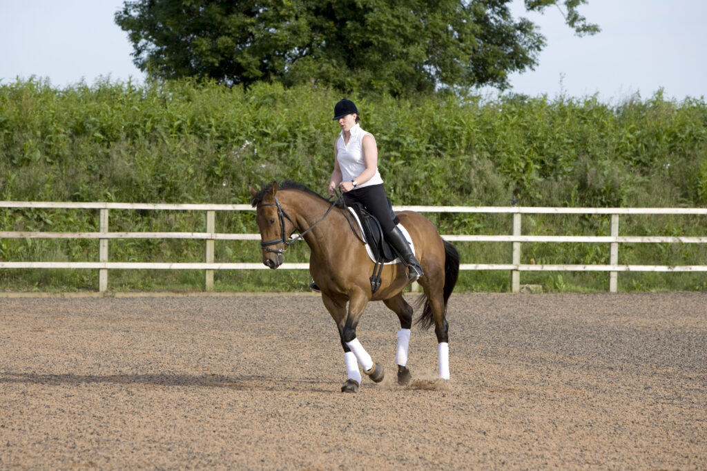
A lameness assessment usually starts with careful observation at walk and trot, often on a firm, level surface. Your vet is looking at stride length, rhythm, straightness and how evenly your horse loads and pushes off each limb. They’ll often watch from the side and from behind, because different angles show different clues.
Then they’ll add context. That might mean circles, different surfaces or seeing the horse under saddle if that’s where the issue shows up. The point isn’t to make the horse look worse. It’s to create the conditions where subtle issues are easier to spot, so your vet can start narrowing things down.
“Measuring lameness isn’t about proving anything. It’s about building a clearer picture so you and your vet can make better decisions. The better the picture, the calmer the plan tends to be.”
Chrysanthi Pitaouli, DVM, CertAVP
3. Grading scales: a shared language, not a label
To make lameness easier to describe, vets often use a grading scale. You might hear the AAEP 0–5 scale mentioned, where 0 is not perceptible and 5 is non weight bearing. The middle grades are where many “not quite right” horses sit. They might look fine some of the time, then show it on a circle, on a hard surface, when they’re tired or when they’re ridden.
A grade isn’t a judgement on you or your horse. It’s simply a way of saying “today, in these conditions, this is how clear it is”, so it can be compared later.
4. Making it repeatable: conditions matter
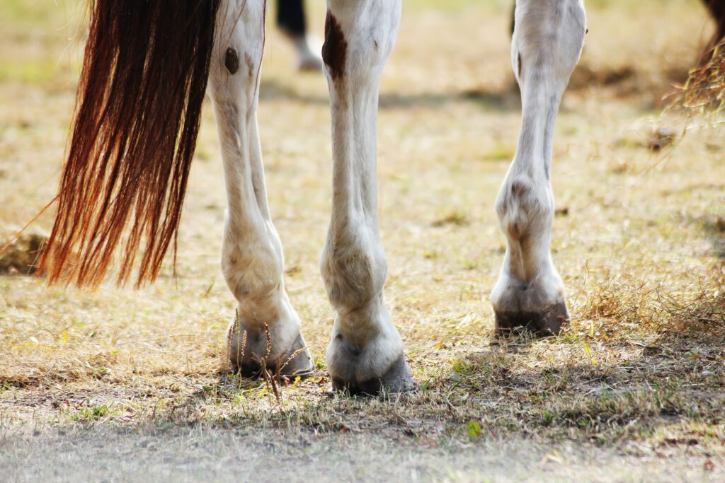
One reason lameness feels so confusing is that it can change depending on:
- the surface (hard vs soft)
- straight lines vs circles
- warming up vs fatigue
- ridden vs in-hand
So part of measuring lameness is keeping things consistent. Vets do this by repeating the same checks and comparing what they see in different situations.
As an owner, you don’t need to recreate a full work-up at home. But you can help by keeping your horse’s story tidy. Two short videos filmed in a similar way a few days apart, plus a note of when it looks better or worse, can be genuinely useful.
5. Objective gait analysis: when sensors help
Alongside the vet’s eye, there are tools designed to measure movement more objectively. Some practices may use systems that allow for small sensors to be placed on areas like the poll and pelvis to measure asymmetry and track change over time.
This can be helpful when:
- signs are subtle and you want to be sure a change is real
- the picture is complicated (for example, multiple issues or compensation)
- your vet wants a consistent way to monitor response to treatment or rehab
It’s worth saying: sensors don’t diagnose the cause on their own. They’re another way of measuring what’s happening, so decisions aren’t based on guesswork.
6. Measuring response: what changes after a test?
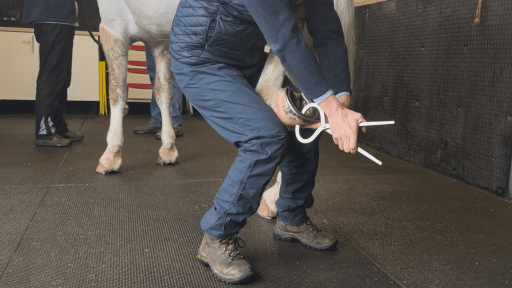
A big part of lameness measurement is comparison. Your vet will look at your horse’s movement at baseline, then see what changes after specific steps in the exam.
That might include flexion tests, hoof testing or local anaesthetic blocks used to narrow down where discomfort is coming from. The principle is the same each time: they’re not just looking at what you see, they’re looking at what changes when something is tested.
This step-by-step approach is why lameness work-ups can feel slow. They’re designed to build accuracy, not rush to a conclusion.
7. Imaging: getting a clearer picture inside
Sometimes the lameness is measurable, but the why still isn’t clear. That’s where imaging comes in.
X-rays and ultrasound can be really useful depending on what your vet suspects. Ultrasound is often used to look at soft tissues that are accessible from the outside. X-rays help with bony changes and joint spaces.
MRI is another option, particularly in complex areas like the foot and lower limb, where structures are crowded and harder to fully assess with other imaging. Standing equine MRI can often be done with the horse standing under mild sedation. It’s one tool among several, used when your vet needs more detail to guide treatment, rehab or next steps.
The takeaway
Measuring lameness isn’t about proving anything. It’s about building a clearer picture so you and your vet can make better decisions. The better the picture, the calmer the plan tends to be.
If you want a practical next step, keep it simple:
- film a short straight line trot clip if it’s safe
- jot down when it looks worse or better (surface, rein, warm-up, ridden vs in-hand)
- share that with your vet
You’re not expected to have the answers. You’re just helping tell your horse’s story clearly and with confidence!