Imaging Explained explores the different modalities available to help with lameness diagnosis, and what you can expect from each. Here we give you the low down on Magnetic Resonance Imaging (MRI).
What is MRI?
Magnetic Resonance Imaging (MRI) is an advanced, non-invasive imaging modality. Considered the gold standard for assessing soft tissue injuries, it is also extremely useful for identifying bone pathology – particularly early change – which can be missed on X-ray. MRI provides valuable information about a lesion’s pathological status and severity, even when conventional radiographs or ultrasound are inconclusive. It is perfectly safe with specialised low-field systems allowing for imaging the distal limb, up to and including the hock and carpus, in a standing, sedated horse.
MRI uses strong magnetic fields and radiofrequency pulses (not X-rays) to generate high-resolution, cross-sectional images. It produces slice-by-slice images in multiple planes, allowing detailed evaluation of both bone and soft tissue structures. Different imaging sequences enable detection of fluid within tissues and characterisation of lesions, which is particularly important for identifying active pathology. In horses, MRI can be performed in the standing, sedated patient for distal limb imaging, allowing assessment under load-bearing conditions, or under general anaesthesia depending on the region and system used.
You can watch a video of how the standing MRI process works for your horse:
When might MRI be used?
- When lameness has been localised to a region but initial imaging (radiography/ultrasound) is inconclusive (most commonly for investigation of foot lameness)
- Following trauma, to assess the structures involved (e.g. penetrating foot injuries)
- To further characterise lesions identified on other imaging modalities (common when soft tissues are involved)
- To monitor lesion progression and response to treatment
What it shows well
- Soft tissues: identification of lesions and characterisation of their severity and activity (tendons, ligaments and supporting structures)
- Bone: structural changes (e.g. sclerosis, resorption/lysis, fractures) and bone oedema-like signal
- Joints: assessment of joint structures, including synovial components; cartilage evaluation may be possible depending on the system
- Injuries where multiple tissue types are involved, MRI helps build a complete picture of the problem area
What it doesn’t show well
- Whole‑horse coverage; it’s targeted to the problem area
- Cartilage assessment can be limited and not always possible – cartilage is a difficult thing to assess with all imaging modalities
- Fibre pattern of tendon lesions
- Hoof capsule and solar bruising
What to expect from MRI
Standing low field MRI allows scanning the lower limb under mild sedation. However, high field systems (often used for heads or higher limb regions) may require general anaesthesia, depending on the centre. Standing MRI is safe and effective with just mild sedation and has been proved diagnostic in over 90% of lameness cases [1].
Regardless of system used, MRI commonly takes longer than other imaging methods (X-ray or ultrasound). Careful examination to localise the suspected problem area before performing MRI is advised.
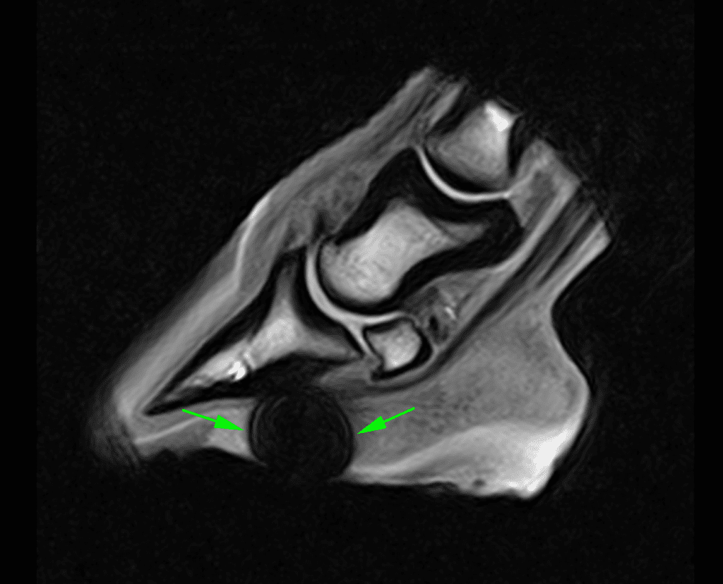
This MRI image clearly shows where a rusty nail had penetrated deep into the horse’s foot. The green arrows show artefact (or distortion) from metal fragments.
Where it fits into the bigger picture
MRI is often the advanced test of choice once lameness is localised, but not explained by first‑line imaging such as X-ray and ultrasound. MRI is especially for structures inside the hoof capsule and now – with advances in technology – standing MRI can scan higher up the limb to aid a definitive diagnosis in cases that were once deemed too tricky. With standing MRI making such a difference to the outcome, you have to ask “Why don’t more horse owners think of MRI for lameness?“.
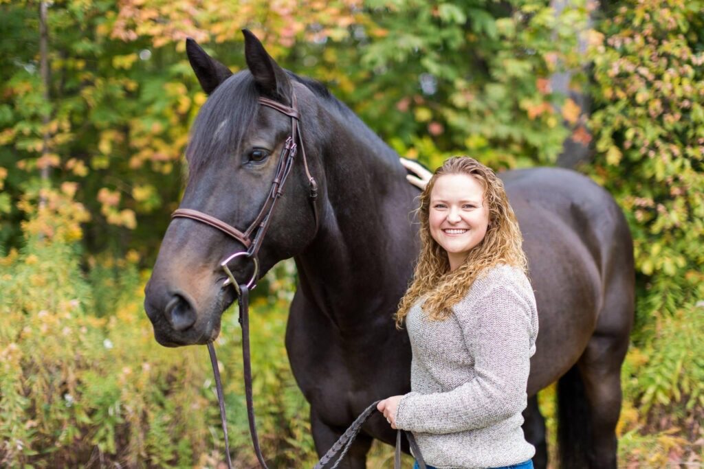
The bottom line is, lack of awareness. So, the more you know, the more likely you are to ask for what you want. Just as Ellie did…
Find out how imaging with standing MRI made the difference to Berkeley as owner Ellie shares her lameness story. No spoiler alerts here but – to quote Ellie:
I got the MRI results in less than 24 hours and had the answers I needed.”
Ellie Woznica