Imaging Explained explores the different modalities available to help with lameness diagnosis and what you can expect from each. Let’s take a look at X-ray.
What is X-ray?
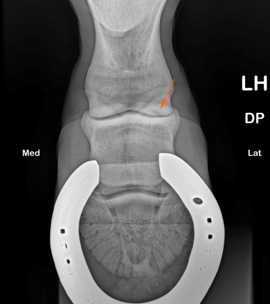
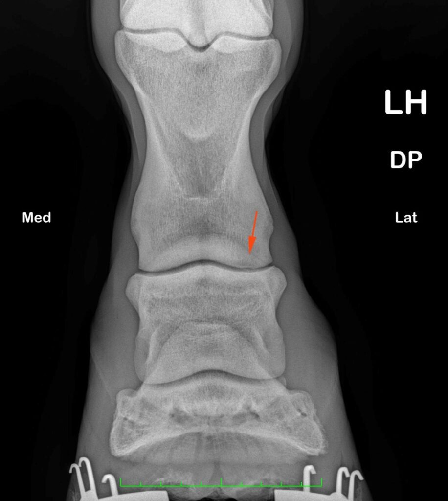
X-ray, or digital radiography, is one of the primary imaging modalities, mainly used for assessing bone injuries and detecting things like fractures, arthritis or bone ‘chips’. A major benefit is that it is often portable (ambulatory) and generally affordable. However, X-rays are limited by the fact that they produce 2D images with superimposition of overlying structures. This limitation can make interpretation of complex areas difficult – for example the hock joint – even with multiple X-rays taken from different angles. In addition, they do not provide information for the soft tissues, meaning concurrent soft tissue injuries may go undetected.
When might we use X-ray?
If your vet suggests a quick look at your horse’s bones and joints, X-rays are often the first step. They are quick, affordable and widely available. They are particularly useful for assessing bone, including fractures, changes in bone density (such as sclerosis or lysis) and new bone formation. However, X-rays provide limited information on soft tissues such as tendons and ligaments, so vets often pair them with ultrasound or, if needed, advanced imaging. It’s a step by step process!
- Sudden, significant lameness after a knock or suspected fracture
- Ongoing joint discomfort or suspected arthritis
- Assessing bone and joint structures after injury, including monitoring healing or progression over time
What it shows well
- Bone: fractures, bone fragments (chips), changes in bone density (sclerosis or lysis), cystic lesions, and new bone formation (including at joint margins or ligament/tendon attachments)
- Joints: changes such as joint space narrowing, remodelling, and new bone formation around the joint
- Overall bone shape and structure in the area imaged
What it doesn’t show
- Soft tissues (e.g., tendons/ligaments) are poorly visualised
- Early bone injury, bone marrow change and subtle or incomplete fractures
- Advanced imaging may be needed if signs don’t match X-ray findings
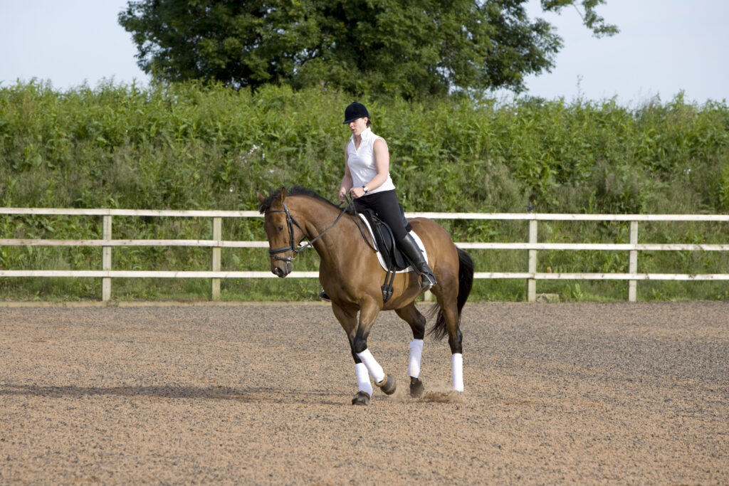
What to expect from X-ray
Portable digital X‑ray units are common in ambulatory practice. Most horses struggling with lameness can be imaged standing, and at home in the yard or field. Your vet will position a plate behind the leg and briefly activate the X‑ray source. If needed, sedation is sometimes used to help your horse stand quietly for the procedure.
Where it fits into the bigger picture
X‑rays are a cornerstone of lameness work‑ups. However, if they fail to fully explain the cause of lameness, your vet may recommend further investigation with CT or MRI. Complementary to each other, they will help provide more detail.